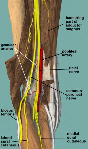
Menisci cover approximately 2/3 of your tibia surface and are thinner on the inside and thicken toward the outer peripheral. They are made of a dense, collagen connective tissue that is tougher than articular cartilage, called fibro-cartilage. The menisci are crescent shaped wedges located in the knee joint at the bottom of your thigh bone and on top of the flat upper surface of your shin bone. A pivot, twist or over-extension of the knee can lead to an ACL strain or tear. The knee ligament that is most frequently injured is the anterior cruciate ligament. The LCL runs along the outside of the knee joint, it provides stability to the lateral (outer) part of the knee.The MCL runs along the inside of the knee joint, it provides stability to the medial (inner) part of the knee.The PCL is in the center of the knee, it limits backward leg movements.The ACL is in the center of the knee, it limits rotation and forward leg movements.There are four ligaments in the knee joint that connect the femur and tibia the anterior cruciate ligament (ACL), the posterior cruciate ligament (PCL), the medial collateral ligament (MCL), and the lateral collateral ligament (LCL). Ligaments are strong, elastic-like tissues that connect bone to bone and provide stability and protection to your knee joint by limiting the forward and backward movement of the shin bone.

This tendon is susceptible to quadriceps tendinitis. The tendon of the quadriceps runs from the quadriceps muscles, down both sides of the patella and join on either side of the tibia. The quadriceps muscles located at the front of your thigh (rectus femoris, vastus lateralis, vastus medialis, vastus intermedius), allow you to straighten your legs and the hamstring muscles, located on the back of your thigh (semitendinosus, semimembranosus, biceps femoris), allow you to bend your knees. The upper leg muscles provide your knees with mobility (extension, flexion and rotation) and strength. The patella tendon is most commonly injured or inflicted with tendonitis, known as Jumper's Knee (patellar tendonitis). It is approximately 4 inches long and inserts at the top of the tibia and spreads over top of the patella where it connects to the quadriceps tendon. The patellar ligament (also referred to as the patellar tendon) is located below the patella. It protects the bones and soft tissue in your knee joint and slides when your knee moves, allowing leverage in your leg muscles.

The patella is the larger sesamoid bone in your body and rests over a groove at the bottom of the femur and the top of the tibia.


Bones embedded in tendons are called sesamoid bones and they protect the tendons and improve the function of the joint by holding the tendons away from the center of the joint. Your patella, or knee cap, is a circular-triangular bone, approximately 2 inches across, that is embedded between the quadriceps tendon above and patellar tendon below. The soft tissue in the knee joint (tendons, ligaments, menisci, cartilage) that provides stability in the knee and hold the bones together at the joint. flexion, extension, medial rotation, and lateral rotation) and it connects the tibia and the fibula, with the thigh bone (femur). It is a pivotal hinge joint in the leg that allows for a variety of movements (i.e. The knee is the largest joint in your body and one of the most easily injured.


 0 kommentar(er)
